Supporting consistent and efficient tissue staining across a range of applications.
Validated on Microfluidic Platform
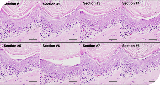
Histology (H&E)
Consistent hematoxylin and eosin staining across multiple tissue sections.
H&E staining of the mouse esophagus Eight sections of the mouse esophagus for H&E staining. Scale bar=50μm
Immunohistochemistry (IHC)
Reproducible immunohistochemical staining with controlled reagent delivery
LYVE1 IHC staining of the mouse stomach Eight sections of the mouse stomach for staining with anti-LYVE1. Scale bar=50μm.
Rapid IHC Demonstration
Microfluidic control enables efficient reagent delivery, thereby reducing incubation times in immunohistochemical staining.
LYVE1 IHC staining of the mouse stomach using a primary antibody incubation of 15 minutes.
Frozen Tissue Section (H&E)
H&E staining was performed on frozen mouse esophagus tissue to evaluate microfluidic staining performance on frozen sections.
The stained sections show clear nuclear detail and well-preserved tissue morphology.
Research Applications
Flexible workflows for experimental studies and method development.
Controlled reagent delivery for sensitive assays
Reduced reagent consumption for valuable reagents
Reproducible conditions across multiple samples
Expanding Capabilities
The microfluidic platform is being extended to support additional staining and analysis methods. Microfluidic control supports exploration of reduced incubation times in immunohistochemistry and evaluation of staining performance on frozen tissue sections.
These capabilities enable method development for rapid staining workflows and comparative studies across sample types.
Immunofluorescence (IF)
Fluorescent Labeling With Controlled Reagent Exposure
In Situ Hybridization (ISH)
Spatial Detection Of Nucleic Acids Within Tissue Sections
TUNEL Assay
Detection of DNA fragmentation in apoptosis studies
Explore Microfluidic Staining in Your Applications
From laboratory workflows to data-driven diagnostics